Chronic Renal Failure and Renal Dialysis

Chronic Renal Failure (CKD) is a worldwide public health problem and is now recognized as a common condition that is associated with an increased risk of cardiovascular disease and chronic renal failure (CRF).
Defines chronic kidney disease as either kidney damage or a decreased kidney glomerular filtration rate (GFR) of less than 60 mL/min/1.73 m2 for 3 or more months.
Whatever the underlying etiology, the destruction of renal mass with irreversible sclerosis and loss of nephrons leads to a progressive decline in GFR.
Classification of the stages of chronic kidney disease, as follows:
Stage 1: Kidney damage with normal or increased GFR (>90 mL/min/1.73 m2)
Stage 2: Mild reduction in GFR (60-89 mL/min/1.73 m2)
Stage 3: Moderate reduction in GFR (30-59 mL/min/1.73 m2)
Stage 4: Severe reduction in GFR (15-29 mL/min/1.73 m2)
Stage 5: Kidney failure (GFR <15>
stage 2 chronic kidney disease, GFR alone does not clinch the diagnosis.
Other markers of kidney damage, including abnormalities in the composition of blood or urine or abnormalities in imaging tests, should also be present in establishing a diagnosis of stage 1 and stage 2 chronic kidney disease. Pathophysiology

Approximately 1 million nephrons are present in each kidney, each contributing to the total GFR. Regardless of the etiology of renal injury, with progressive destruction of nephrons, the kidney has an innate ability to maintain GFR by hyperfiltration and compensatory hypertrophy of the remaining healthy nephrons.
This nephron adaptability allows for continued normal clearance of plasma solutes so that substances such as urea and creatinine start to show significant increases in plasma levels only after total GFR has decreased to 50%, when the renal reserve has been exhausted. The plasma creatinine value will approximately double with a 50% reduction in GFR. A rise in plasma creatinine from a baseline value of 0.6 mg/dL to 1.2 mg/dL in a patient, although still within the reference range, actually represents a loss of 50% of functioning nephron mass.
The residual nephron hyperfiltration and hypertrophy, although beneficial for the reasons noted, has been hypothesized to represent a major cause of progressive renal dysfunction.
Factors other than the underlying disease process and glomerular hypertension that may cause progressive renal injury include the following:
· Systemic hypertension
· Acute insults from nephrotoxins or decreased perfusion
· Proteinuria
· Increased renal ammonia with interstitial injury
· Hyperlipidemia
· Hyperphosphatemia with calcium phosphate deposition
· Decreased levels of nitrous oxide
· Smoking

Frequency in United States
In the United States, there is a rising incidence and prevalence of kidney failure, with poor outcomes and high cost. Kidney disease is the ninth leading cause of death in the United States. Data from the United States Renal Data System (USRDS) indicated that there has been an increase of 104% in the prevalence of chronic renal failure (CRF) between the years 1990-2001. There is an even higher prevalence of the earlier stages of chronic kidney disease.
International prevalence
The incidence rates of end-stage renal disease (ESRD) have increased steadily internationally since 1989. The United States has the highest incident rate of ESRD, followed by Japan. Japan has the highest prevalence per million population, with the United States taking second place.
Mortality/Morbidity
Chronic kidney disease is a major cause of morbidity and mortality, particularly at the later stages. Although the diabetic population is at highest risk, in the United States, the general hemodialysis and peritoneal dialysis populations have 2 hospital admissions per patient per year; patients who have a renal transplant have an average of 1 hospital admission per year. The 5-year survival rate for a patient undergoing chronic dialysis in the United States is approximately 35%.
This is approximately 25% in patients with diabetes. The most common cause of death in the dialysis population is cardiovascular disease.

Among patients with ESRD end-stage renal disease, aged 65 years and older, the mortality rates are 6 times higher than in the general population. In 2003, over 69,000 dialysis patients enrolled in the ESRD program died (annual adjusted mortality rate of 210.7 per 1000 patient-years at risk for the dialysis population, which represents a 14% decrease since peaking at 244.5 per 1000 patient-years in 1988).
The highest mortality rate is within the first 6 months of initiating dialysis, which then tends to improve over the next 6 months, before increasing gradually over the next 4 years.
At age 60 years, a healthy person can expect to live for more than 20 years, whereas the life expectancy of a 60-year-old patient starting hemodialysis is closer to 4 years.
Race
Chronic kidney disease affects all races, but, in the United States, a significantly higher incidence of ESRD exists in blacks as compared to whites; the incident rate for blacks is nearly 4 times that for whites.
Sex
The chronic kidney disease stages was similar in both sexes.
Age
Chronic kidney disease is found in persons of all ages. The biologic process of aging initiates various structural and functional changes within the kidney. Renal mass progressively declines with advancing age. Glomerulosclerosis leads to a decrease in renal weight.
Histologic examination is notable for a decrease in glomerular number of as much as 30-50% by age 70 years.


Ischemic obsolescence of cortical glomeruli is predominant, with relative sparing of the renal medulla. Juxtamedullary glomeruli see a shunting of blood from the afferent to efferent arterioles, resulting in redistribution of blood flow favoring the renal medulla.
These anatomical and functional changes in renal vasculature appear to contribute to an age-related decrease in renal blood flow. Renal hemodynamic measurements in aged human and animals suggest that altered functional response of the renal vasculature may be an underlying factor in diminished renal blood flow and increased filtration noted with progressive renal aging.
CLINICAL investigation of CKD
History
Patients with chronic kidney disease
stages 1-3 (GFR >30 mL/min) are generally asymptomatic and do not experience clinically evident disturbances in water or electrolyte balance or endocrine/metabolic derangements. Generally, these disturbances clinically manifest with chronic kidney disease
stages 4-5 (GFR <30>


 Drug Name
Calcium acetate (Calphron, PhosLo)
 Drug Name
Calcium carbonate (Caltrate, Oystercal
 Drug Name
Calcitriol (Rocaltrol, Calcijex)

 Drug Name
Sevelamer (Renagel)
 Drug Name
Paricalcitol (Zemplar)

Contraindications
Documented hypersensitivity; uncontrolled hypertension
Pregnancy
C - Fetal risk revealed in studies in animals but not established or not studied in humans; may use if benefits outweigh risk to fetus
Precautions
Caution in porphyria, hypertension, and history of seizures; decrease dose if hematocrit increase exceeds 4 U in any 2-wk period

Drug Name
Ferrous sulfate (Feosol, Feratab, Slow FE)
Description
Used as a building block for hemoglobin synthesis in treating anemia of chronic kidney disease with erythropoietin.
Adult Dose
325 mg PO qd/tid

Contraindications
Documented hypersensitivity; iron overload; anemia unrelated to iron deficiency
Interactions
Decreases bioavailability of orally administered iron
Pregnancy
B - Fetal risk not confirmed in studies in humans but has been shown in some studies in animals
Precautions
May cause hypotension (related to IV administration rate or cumulative dose), cramps, headache, nausea, vomiting, diarrhea, or anaphylaxis

These anatomical and functional changes in renal vasculature appear to contribute to an age-related decrease in renal blood flow. Renal hemodynamic measurements in aged human and animals suggest that altered functional response of the renal vasculature may be an underlying factor in diminished renal blood flow and increased filtration noted with progressive renal aging.
CLINICAL investigation of CKD
History
Patients with chronic kidney disease
stages 1-3 (GFR >30 mL/min) are generally asymptomatic and do not experience clinically evident disturbances in water or electrolyte balance or endocrine/metabolic derangements. Generally, these disturbances clinically manifest with chronic kidney disease
stages 4-5 (GFR <30>

The ability to maintain potassium (K) excretion at near normal levels is generally maintained in chronic kidney disease patients as long as both aldosterone secretion and distal flow are maintained. Another defense against potassium retention in patients with chronic kidney disease is increased potassium excretion in the GI tract, which also is under control of aldosterone.
Therefore, hyperkalemia usually develops when the GFR falls to less than 20-25 mL/min because of the decreased ability of the kidneys to excrete potassium. It can be observed sooner in patients who ingest a potassium-rich diet or if serum aldosterone levels are low, such as in type IV renal tubular acidosis commonly observed in people with diabetes or with use of angiotensin-converting enzyme (ACE) inhibitors or nonsteroidal anti-inflammatory drugs (NSAIDs).
Therefore, hyperkalemia usually develops when the GFR falls to less than 20-25 mL/min because of the decreased ability of the kidneys to excrete potassium. It can be observed sooner in patients who ingest a potassium-rich diet or if serum aldosterone levels are low, such as in type IV renal tubular acidosis commonly observed in people with diabetes or with use of angiotensin-converting enzyme (ACE) inhibitors or nonsteroidal anti-inflammatory drugs (NSAIDs).

Hyperkalemia in chronic kidney disease can be aggravated by an extracellular shift of potassium, such as that occurs in the setting of acidemia or from lack of insulin. Hypokalemia is uncommon but can develop among patients with very poor intake of potassium, gastrointestinal or urinary loss of potassium, diarrhea, or diuretic use.
Metabolic acidosis has been shown to have deleterious effects on protein balance, leading to a negative nitrogen balance, increased protein degradation, increased essential amino acid oxidation, reduced albumin synthesis, and a lack of adaptation to a low protein diet. Hence, this is associated with protein-energy malnutrition, loss of lean body mass, and muscle weakness. The mechanism for reducing protein may include effects on ATP-dependent ubiquitin proteasomes and increased activity of branched chain keto acid dehydrogenases.
Hypoalbuminemia (a marker of protein-energy malnutrition and a powerful predictive marker of mortality in dialysis patients as well as in the general population) was independently associated with low bicarbonate as well as the inflammatory marker C reactive protein.

Metabolic acidosis is a factor in the development of renal osteodystrophy, as bone acts as a buffer for excess acid, with resultant loss of mineral. Acidosis may interfere with vitamin D metabolism, and patients who are persistently more acidotic are more likely to have osteomalacia or low-turnover bone disease.
Salt and water handling by the kidney is altered in patients with chronic kidney disease. Extracellular volume expansion and total-body volume overload results from failure of sodium and free water excretion.
This generally becomes clinically manifested when the GFR falls to less than 10-15 mL/min, when compensatory mechanisms have become exhausted. As kidney function declines further, sodium retention and extracellular volume expansion lead to peripheral and, not uncommonly, pulmonary edema and hypertension. At a higher GFR, excess sodium and water intake could result in a similar picture if the ingested amounts of sodium and water exceed the available potential for compensatory excretion.

Normochromic normocytic anemia principally develops from decreased renal synthesis of erythropoietin, the hormone responsible for bone marrow stimulation for red blood cell (RBC) production. It starts early in the course of disease and becomes more severe as the GFR progressively decreases with the availability of less viable renal mass. RBC survival is decreased, and tendency of bleeding is increased from the uremia-induced platelet dysfunction.
Other causes of anemia in chronic kidney disease patients include chronic blood loss, secondary hyperparathyroidism, inflammation, nutritional deficiency, and accumulation of inhibitors of erythropoiesis.
Anemia is associated with fatigue, reduced exercise capacity, impaired cognitive and immune function, and reduced quality of life. Anemia is also associated with the development of cardiovascular disease, the new onset of heart failure, or the development of more severe heart failure. Anemia is associated with increased cardiovascular mortality.

Renal bone disease is a common complication of chronic kidney disease and results in both skeletal complications (eg, abnormality of bone turnover, mineralization, linear growth) and extraskeletal complications (eg, vascular or soft tissue calcification).
Different types of bone disease occur with chronic kidney disease, as follows:
(1) high turnover bone disease due to high parathyroid hormone (PTH) levels
(2a) low turnover bone disease (adynamic bone disease)
(2b) defective mineralization (osteomalacia)
(3) mixed disease
(4) beta-2-microglobulin associated bone disease.
Calcium and calcitriol are primary feedback inhibitors; hyperphosphatemia is a stimulus to PTH synthesis and secretion.
Phosphate retention begins in early chronic kidney disease; when the GFR falls, less phosphate is filtered and excreted, but serum levels do not rise initially because of increased PTH secretion, which increases renal excretion.
As the GFR falls toward chronic kidney disease stages 4-5, hyperphosphatemia develops from the inability of the kidneys to excrete the excess dietary intake.
Low serum calcitriol levels, hypocalcemia, and hyperphosphatemia have all been demonstrated to independently trigger PTH synthesis and secretion.
If serum levels of PTH (parathyroid hormone) remain elevated, a high bone turnover lesion, known as osteitis fibrosa, develops. This is one of several bone lesions, which as a group are commonly known as renal osteodystrophy. These lesions develop in patients with severe chronic kidney disease and are common in those with ESRD.

osteitis fibrosa
The prevalence of adynamic bone disease has increased, and it has been described before the initiation of dialysis in some cases. The pathogenesis of adynamic bone disease is not well defined, but several factors may contribute,
· including high calcium load
· use of vitamin D sterols
· increasing age
· previous corticosteroid therapy
· peritoneal dialysis
Other manifestations of uremia in ESRD (End stage renal disease) many of which are more likely in patients who are inadequately dialyzed, include the following:
· Pericarditis - Can be complicated by cardiac tamponade, possibly resulting in death.
· Encephalopathy - Can progress to coma and death
· Peripheral neuropathy
· Restless leg syndrome
· GI symptoms - Anorexia, nausea, vomiting, diarrhea
· Skin manifestations - Dry skin, pruritus, ecchymosis
· Fatigue, increased somnolence, failure to thrive
· Malnutrition
· Erectile dysfunction, decreased libido, amenorrhea
· Platelet dysfunction with tendency to bleeding
Physical examination
Often is not very helpful but may reveal findings characteristic of the condition underlying chronic kidney disease (eg, lupus, severe arteriosclerosis, hypertension) or complications of chronic kidney disease (eg, anemia, bleeding diathesis, pericarditis).
Causes of CRF.
· Vascular disease - Renal artery stenosis, atheroemboli, hypertensive nephrosclerosis, renal vein thrombosis
· Primary glomerular disease - Membranous nephropathy, immunoglobulin A (IgA) nephropathy, minimal change disease, rapidly progressive (crescentic) glomerulonephritis
· Secondary glomerular disease - Diabetes mellitus, systemic lupus erythematosus, rheumatoid arthritis, mixed connective tissue disease, scleroderma, postinfectious glomerulonephritis, endocarditis, hepatitis B and C, syphilis, human immunodeficiency virus (HIV), parasitic infection, heroin use, gold, penicillamine, amyloidosis.
· Tubulointerstitial disease - Drugs (eg, sulfa, allopurinol), infection (viral, bacterial, parasitic), chronic hypokalemia, chronic hypercalcemia, heavy metals, radiation nephritis, polycystic kidneys.
· Urinary tract obstruction - Urolithiasis, benign prostatic hypertrophy, tumors, retroperitoneal fibrosis, urethral stricture, neurogenic bladder
Lab Studies

· Serum electrolytes, BUN, and creatinine - The BUN and creatinine levels will be elevated in patients with chronic kidney disease. Hyperkalemia or low bicarbonate levels may be present in patients with chronic kidney disease.
· Serum calcium, phosphate, vitamin D, and intact parathyroid hormone (PTH) levels are obtained to look for evidence of renal bone disease.
· CBC count - Normochromic normocytic anemia is commonly seen in chronic kidney disease. Other underlying causes of anemia should be ruled out.
· Serum albumin - Patients may have hypoalbuminemia due to urinary protein loss or malnutrition.
· Lipid profile - A lipid profile should be performed in all patients with chronic kidney disease because of their increased risk of cardiovascular disease.
· Urinalysis - Dipstick proteinuria may suggest a glomerular or tubulointerstitial problem. The urine sediment finding of RBCs, RBC casts, suggests proliferative glomerulonephritis. Pyuria and/or WBC casts are suggestive of interstitial nephritis (particularly if eosinophiluria is present) or urinary tract infection.
· Spot urine collection for total protein-to-creatinine ratio allows reliable approximation (extrapolation) of total 24-hour urinary protein excretion. A value of greater than 2 g is considered to be within the glomerular range, and a value of greater than 3-3.5 g is within the nephrotic range; less than 2 is characteristic of tubulointerstitial problems.
· Twenty-four–hour urine collection for total protein and CrCl
In certain cases, the following tests may be ordered as part of the evaluation of patients with chronic kidney disease:
· Serum and urine protein electrophoresis to screen for a monoclonal protein possibly representing multiple myeloma
· Antinuclear antibodies (ANA), double-stranded DNA antibody levels to screen for systemic lupus erythematosus
· Serum complement levels.
Imaging Studies

· Plain abdominal x-ray - Particularly useful to look for radio-opaque stones or nephrocalcinosis
· Intravenous pyelogram - Not commonly used because of potential for intravenous contrast renal toxicity; often used to diagnose renal stones
· Renal ultrasound - Small echogenic kidneys are observed in advanced renal failure..
· Renal radionuclide scan - Useful to screen for renal artery stenosis when performed with captopril administration but is unreliable for GFR of less than 30 cc/min; also quantitates differential renal contribution to total GFR
· CT scan - CT scan is useful to better define renal masses and cysts usually noted on ultrasound. Also, it is the most sensitive test for identifying renal stones.
· MRI is very useful in patients who require a CT scan but who cannot receive intravenous contrast. It is reliable in the diagnosis of renal vein thrombosis, as are CT scan and renal venography.
· Magnetic resonance angiography also is becoming more useful for diagnosis of renal artery stenosis, although renal arteriography remains the criterion standard.
· Voiding cystourethrogram (VCUG) - Criterion standard for diagnosis of vesicoureteral reflux
Other Tests
The Cockcroft-Gault formula for estimating CrCl (Creatinine clearance tests) should be used routinely as a simple means to provide a reliable approximation of residual renal function in all patients with chronic kidney disease. The formulas are as follows:
· CrCl (male) = ([140-age] X weight in kg)/(serum creatinine X 72)
· CrCl (female) = CrCl (male) X 0.85
Percutaneous renal biopsy is performed most often with ultrasound guidance and the use of a mechanical gun.
Surgical open renal biopsy can be considered when the risk of renal bleeding is felt to be great, occasionally with solitary kidneys, or when percutaneous biopsy is technically difficult to perform.
Histologic Findings
Renal histology in chronic kidney disease reveals findings compatible with the underlying primary renal diagnosis.
Medical Care
The medical care of patients with chronic kidney disease should focus on the following:
· Delaying or halting the progression of chronic kidney disease
· Treatment of the underlying condition if possible
· Aggressive blood pressure control to target values per current guidelines. Systolic blood pressure control is considered more important and is also considered difficult to control in elderly patients with chronic kidney disease.
· Use of ACE inhibitors or angiotensin receptor blockers as tolerated, with close monitoring for renal deterioration and for hyperkalemia
· Data support the use of ACE inhibitors/angiotensin receptor blockers in diabetic kidney disease with or without proteinuria. However, in nondiabetic kidney disease, ACE inhibitors/angiotensin receptor blockers are effective in retarding the progression of disease among patients with proteinuria of less of than 500 mg/d.
· Aggressive glycemic control per the American Diabetes Association (ADA) recommendations (target HbA1C <7%) name="TreatmentConsultations">
Consultations
· Early nephrology referral (decreases morbidity and mortality)
· Renal dietitian
· Vascular surgery for permanent vascular access
· General surgery for peritoneal catheter placement
· Referral to renal transplant center
· Diet
· Protein restriction early in chronic kidney disease as a means to delay a decline in the GFR is controversial; however, as the patient approaches chronic kidney disease stage 5, this is recommended to delay the onset of uremic symptoms.
· Patients with chronic kidney disease who already are predisposed to becoming malnourished are at higher risk for malnutrition with overly aggressive protein restriction.
· Malnutrition is a well-established predictor of increased morbidity and mortality in the ESRD population and must be avoided if possible.
· Phosphate restriction starting early in chronic kidney disease
· Potassium restriction
· Sodium and water restriction as needed to avoid volume overload
Drug Category: Phosphate-lowering agents
· Hyperphosphatemia is treated with dietary phosphate binders and dietary phosphate restriction.
· Hypocalcemia is treated with calcium supplements and possibly calcitriol.
· Hyperparathyroidism is treated with calcitriol or vitamin D analogs.
 Drug Name
Calcium acetate (Calphron, PhosLo)
Description
For treatment of hyperphosphatemia in chronic kidney disease. Combines with dietary phosphorus to form insoluble calcium phosphate, which is excreted in feces.
Adult Dose
1334 mg PO with each meal; increase to bring serum phosphate value to 6 mg/dL as long as hypercalcemia does not develop; may require as much as 2668 mg
Contraindications
Hypercalcemia; hypophosphatemia; renal calculi
Interactions
May increase effect of quinidine; may decrease effects of tetracyclines, atenolol, salicylates, iron salts, and fluoroquinolones; IV administration antagonizes effects of verapamil; large intakes of dietary fiber may decrease calcium absorption and levels
Pregnancy
C - Fetal risk revealed in studies in animals but not established or not studied in humans; may use if benefits outweigh risk to fetus
Precautions
Hypercalcemia or hypercalcuria may occur when therapeutic amounts are administered
Drug Name
Calcium carbonate (Caltrate, Oystercal
Description
For treatment of hyperphosphatemia or as a calcium supplement in chronic kidney disease. Successfully normalizes phosphate concentrations in patients with chronic kidney disease. Combines with dietary phosphate to form insoluble calcium phosphate, which is excreted in feces. Marketed in a variety of dosage forms and is relatively inexpensive.
Adult Dose
1-2 g PO divided bid/qid; with meals as a phosphorous binder; between meals as a calcium supplement
Pediatric Dose
45-65 mg/kg/d PO divided qid
Contraindications
Renal calculi; hypercalcemia; hypophosphatemia; digitalis toxicity
Pregnancy
C - Fetal risk revealed in studies in animals but not established or not studied in humans; may use if benefits outweigh risk to fetus
Precautions
Hypercalcemia or hypercalcuria may occur when therapeutic amounts are administered
 Drug Name
Calcitriol (Rocaltrol, Calcijex)
Description
Used to suppress parathyroid production and secretion in secondary hyperparathyroidism and for treatment of hypocalcemia in chronic kidney disease by increasing intestinal calcium absorption.
Adult Dose
0.25 mcg PO qd/qod0.5 mcg IV qd 3 times/wkIncrease at 4- to 8-wk intervals by 0.25-mcg/d to achieve target PTH level and to maintain serum calcium levels at 9-10 mg/dL
Pediatric Dose
Initial: 15 ng/kg/d POMaintenance: 5-40 ng/kg/d PO
Contraindications
Documented hypersensitivity; hypercalcemia; malabsorption syndrome
Interactions
Cholestyramine and colestipol decrease absorption; magnesium-containing antacids and thiazide diuretics can increase calcitriol effects
Pregnancy
C - Fetal risk revealed in studies in animals but not established or not studied in humans; may use if benefits outweigh risk to fetus
Precautions
Adequate response to calcitriol in improving hypocalcemia depends on adequate dietary calcium intake; serum calcium phosphate product must not exceed 70 mg/dL to minimize metastatic tissue and blood vessel calcification; avoid hypercalcemia

Drug Name
Doxercalciferol (Hectorol)
Description
A vitamin D analog (1-alpha-hydroxyergocalciferol) that does not require activation by the kidneys. Indicated for the treatment of secondary hyperparathyroidism in end-stage renal disease.
Adult Dose
10 mcg PO 3 times/wk at dialysis; adjust dose as needed to lower blood iPTH to 150-300 pg/mL; increase dose by 2.5 mcg/8 wk if iPTH is not lowered by 50% and fails to reach the target range; not to exceed 20 mcg/3 times/wkAlternatively, 4 mcg IV 3 times/wk; may adjust dose by 1-2 mcg/8 wk to maintain iPTH levels
Contraindications
Documented hypersensitivity; recent hypercalcemia or hyperphosphatemia
Pregnancy
B - Fetal risk not confirmed in studies in humans but has been shown in some studies in animals
Precautions
May cause headache, malaise, dyspnea, or hypercalcemia; caution in renal osteodystrophy with hyperphosphatemia (potential for metastatic calcification)
 Drug Name
Lanthanum carbonate (Fosrenal)
Description
Noncalcium, nonaluminum phosphate binder indicated for reduction of high phosphorus levels in patients with end-stage renal disease. Directly binds dietary phosphorus in upper GI tract, thereby inhibiting phosphorus absorption.
Adult Dose
Initial: 250-500 mg PO tid pc (chewable tabs); adjust dose q2-3wk to target serum phosphorus levelMaintenance: 500-1000 mg PO tid pc
Pediatric Dose
Not established
Contraindications
Documented hypersensitivity; bowel obstruction; hypophosphatemia
Interactions
Drugs known to interact with antacids (eg, alendronate, amprenavir, ciprofloxacin, itraconazole, tetracycline, thyroid hormones) should not be administered within 2 h
Pregnancy
C - Fetal risk revealed in studies in animals but not established or not studied in humans; may use if benefits outweigh risk to fetus
Precautions
Deposited into developing bone, including growth plate (long-term effects unknown); common adverse effects typically diminish over time but include headache, abdominal pain, nausea, diarrhea, constipation, and vomiting.
 Drug Name
Sevelamer (Renagel)
Description
Indicated for the reduction of serum phosphorous in patients with ESRD. Binds dietary phosphate in the intestine, thus inhibiting its absorption. In patients on hemodialysis, it decreases the frequency of hypercalcemic episodes relative to patients on calcium acetate treatment.
Adult Dose
Initial: 800-1600 mg PO tid with mealsMaintenance: Increase or decrease by 400-800 mg per meal q2wk to maintain serum phosphorous at 6 mg/dL or less
Contraindications
Documented hypersensitivity; bowel obstruction; hypophosphatemia
Pregnancy
C - Fetal risk revealed in studies in animals but not established or not studied in humans; may use if benefits outweigh risk to fetus
Precautions
Caution in patients with dysphagia, severe GI tract motility disorders, or swallowing disorders; does not contain calcium or alkali supplementation (monitor serum calcium, bicarbonate, and chloride levels)
 Drug Name
Paricalcitol (Zemplar)
Description
For treatment of secondary hyperparathyroidism in ESRD. Reduces PTH levels, stimulates calcium and phosphorous absorption, and stimulates bone mineralization.
Adult Dose
0.04-0.1 mcg IV bolus 3 times/wk; adjust dose based on PTH levels
Contraindications
Documented hypersensitivity; hypercalcemia; vitamin D toxicity
Interactions
Do not use phosphate or vitamin D-related compounds concomitantly with paricalcitol; caution if administered with digoxin (digitalis toxicity is potentiated by hypercalcemia)
Pregnancy
C - Fetal risk revealed in studies in animals but not established or not studied in humans; may use if benefits outweigh risk to fetus
Precautions
Caution in breastfeeding; adverse effects include GI tract distress, dry mouth, lightheadedness, edema, chills, or fever

Drug Category: Growth factors
Used to treat anemia of chronic kidney disease by stimulating RBC production.
Drug Name
Epoetin alfa (Epogen, Procrit)
Description
Stimulates division and differentiation of committed erythroid progenitor cells. Induces release of reticulocytes from bone marrow into blood stream.
Used to treat anemia of chronic kidney disease by stimulating RBC production.
Drug Name
Epoetin alfa (Epogen, Procrit)
Description
Stimulates division and differentiation of committed erythroid progenitor cells. Induces release of reticulocytes from bone marrow into blood stream.
Adult Dose
50-150 U/kg IV/SC 3 times/wk
50-150 U/kg IV/SC 3 times/wk
Contraindications
Documented hypersensitivity; uncontrolled hypertension
Pregnancy
C - Fetal risk revealed in studies in animals but not established or not studied in humans; may use if benefits outweigh risk to fetus
Precautions
Caution in porphyria, hypertension, and history of seizures; decrease dose if hematocrit increase exceeds 4 U in any 2-wk period

Drug Category: Iron salts
Nutritionally essential inorganic substances used to treat anemia.
Nutritionally essential inorganic substances used to treat anemia.
Drug Name
Ferrous sulfate (Feosol, Feratab, Slow FE)
Description
Used as a building block for hemoglobin synthesis in treating anemia of chronic kidney disease with erythropoietin.
Adult Dose
325 mg PO qd/tid

Drug Name
Iron dextran (DexFerrum, InFed)
Iron dextran (DexFerrum, InFed)
Description
Used to treat microcytic, hypochromic anemia resulting from iron deficiency when oral administration is unfeasible or ineffective.Utilized to replenish iron stores in individuals on erythropoietin therapy who cannot take or tolerate oral iron supplementation.A 0.5-mL (0.25 mL in children) test dose should be administered prior to starting therapy.Available as 50 mg iron/mL (as dextran).
Adult Dose
>50 kg: 100 mg IV (2 mL); not to exceed 2 mL/d
Pediatric Dose
5-10 kg: 50 mg IV (1 mL)
Contraindications
Documented hypersensitivity; anemias that are not involved with iron deficiency; hemochromatosis; hemolytic anemia; acute phase of infectious kidney disease
Interactions
Chloramphenicol-induced bone marrow toxicity may cause increased iron levels
Pregnancy
C - Fetal risk revealed in studies in animals but not established or not studied in humans; may use if benefits outweigh risk to fetus
Precautions
Monitor for allergic reactions (eg, flushing, hypotension, nausea
Drug Name
Iron sucrose (Venofer)
Iron sucrose (Venofer)
Description
Used to treat iron deficiency (in conjunction with erythropoietin) due to chronic hemodialysis. Iron deficiency is caused by blood loss during the dialysis procedure, increased erythropoiesis, and insufficient absorption of iron from the GI tract. Iron sucrose has shown a lower incidence of anaphylaxis than other parenteral iron products.
Adult Dose
5 mL (100 mg elemental iron) IV by slow injection or infusion during dialysis session; typically requires a minimum cumulative dose of 1000 mg of elemental iron over 10 consecutive dialysis sessions to achieve a favorable hemoglobin or hematocrit response; not to exceed 3 doses per wk
Contraindications
Documented hypersensitivity; iron overload; anemia unrelated to iron deficiency
Interactions
Decreases bioavailability of orally administered iron
Pregnancy
B - Fetal risk not confirmed in studies in humans but has been shown in some studies in animals
Precautions
May cause hypotension (related to IV administration rate or cumulative dose), cramps, headache, nausea, vomiting, diarrhea, or anaphylaxis

Drug Category: Recombinant Human Erythropoietin
Stimulates development of erythroid progenitor cells.
Stimulates development of erythroid progenitor cells.
Drug Name
Darbepoetin (Aranesp)
Darbepoetin (Aranesp)
Description
Erythropoiesis stimulating protein closely related to erythropoietin, a primary growth factor produced in kidney that stimulates development of erythroid progenitor cells. Mechanism of action is similar to that of endogenous erythropoietin, which interacts with stem cells to increase red cell production. Differs from epoetin alfa (recombinant human erythropoietin) in containing 5 N-linked oligosaccharide chains, whereas epoetin alfa contains 3. Has longer half-life than epoetin alfa (may be administered weekly or biweekly).
Adult Dose
0.45 mcg/kg IV/SC qwk initially; adjust dose (not to exceed 3 mcg/kg/wk) or frequency (eg, q2wk); to maintain target Hgb (not to exceed 12 g/dL); do not increase dose more frequently than qmoSwitching from epoetin alfa: Base dose on total weekly erythropoietin dose and frequency of administration
Contraindications
Documented hypersensitivity; uncontrolled hypertension
Pregnancy
C - Fetal risk revealed in studies in animals but not established or not studied in humans; may use if benefits outweigh risk to fetus
Precautions
Elevation in Hgb > 1 g/dL/2wk increases risk of MI, neurologic events (eg, seizures, stroke) and exacerbations of hypertension, CHF, thrombosis, ischemia, and edema; adverse effects include infection, hypertension, hypotension, myalgia, headache, and diarrhea (some of adverse events may be due to chronic renal failure or dialysis); severe skin rash may occur (rare)
Drug Category: Calcimimetic agents
These agents reduce parathyroid hormone levels.
These agents reduce parathyroid hormone levels.
Drug Name
Cinacalcet (Sensipar)
 Description
Directly lowers intact parathyroid hormone (iPTH) levels by increasing sensitivity of calcium sensing receptor on chief cell of parathyroid gland to extracellular calcium. Also results in concomitant serum calcium decrease. Indicated for secondary hyperparathyroidism in patients with chronic kidney disease on dialysis.
Adult Dose
30 mg PO qd initially; titrate upward slowly (no more frequent than q2-4wk intervals) by 30 mg increments to target iPTH of 150-300 pg/mL
Take with meals or immediately following; do not crush, chew or cut tablets
Interactions
Strong CYP450 2D6 inhibitor; may increase serum levels of CYP 2D6 substrates (eg, flecainide, vinblastine, thioridazine, tricyclic antidepressants); coadministration with CYP450 3A4 inhibitors (eg, ketoconazole, erythromycin, itraconazole) may decrease cinacalcet clearance
Pregnancy
C - Fetal risk revealed in studies in animals but not established or not studied in humans; may use if benefits outweigh risk to fetus
Precautions
Serum calcium reduction may cause lowered seizure threshold, paresthesia, myalgia, cramping, and tetany; monitor calcium and phosphorus levels closely within 1 wk following initial dose or dose changes, and then monthly (secondary hyperparathyroidism) and q2 mo (parathyroid carcinoma); do not initiate treatment if serum calcium below 8.4 mg/dL; adynamic bone disease may occur if iPTH levels suppressed below 100 pg/mL; caution with hepatic impairment; common adverse effects include nausea and vomiting
FOLLOW-UP
Further Inpatient Care
Patients who develop potentially life-threatening complications of chronic kidney disease should be hospitalized and closely monitored.
Further Outpatient Care
Patients with chronic kidney disease should be referred to a nephrologist early in the course of their disease and have continued nephrologic follow-up until initiation of chronic renal replacement therapy.
A multidisciplinary approach to care, including involvement of the nephrologist, primary care physician, renal dietitian, nurse, and social worker, should be initiated early in the course of chronic kidney disease, with close patient follow-up.
Transfer
Patients with chronic kidney disease acutely presenting with indications for dialytic therapy should be transferred to a hospital center where acute dialysis can be performed.
Prognosis
Patients with chronic kidney disease generally progress to ESRD. The rate of progression depends on the underlying diagnosis, on the successful implementation of secondary preventative measures, and on the individual patient.
Patients on chronic dialysis have a high incidence of morbidity and mortality.
Patients with ESRD who undergo renal transplantation survive longer than those on chronic dialysis.
Patient Education
Patients with chronic kidney disease should be educated about the importance of compliance with secondary preventative measures, natural disease progression, prescribed medications (highlighting their potential benefits and adverse effects), avoidance of nephrotoxins, diet, chronic renal replacement modalities, including peritoneal dialysis, hemodialysis, and transplantation, and permanent vascular access options for hemodialysis.
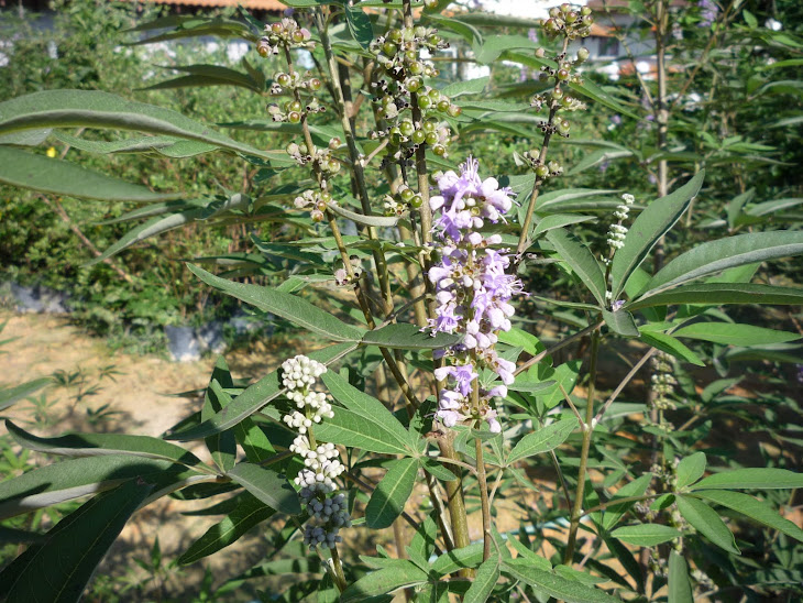
Herbs can help CRF patients on recovery.
1 - Angelica Sinensis
2 - Astragalus mongholicus
3 - Cordyceps sinensis fungus
Please watch the following sites:
http://www.youtube.com/watch?v=Pz5DHAv_Mw4&feature=related
http://www.youtube.com/watch?v=glu0dzK4dbU&feature=related
http://www.youtube.com/watch?v=Kiw-9VvL2Mc&feature=related
http://www.youtube.com/watch?v=dx8wJT9npnE&feature=related
http://www.youtube.com/watch?v=WZosHub0MOQ
Patients with ESRD who undergo renal transplantation survive longer than those on chronic dialysis.
Patient Education
Patients with chronic kidney disease should be educated about the importance of compliance with secondary preventative measures, natural disease progression, prescribed medications (highlighting their potential benefits and adverse effects), avoidance of nephrotoxins, diet, chronic renal replacement modalities, including peritoneal dialysis, hemodialysis, and transplantation, and permanent vascular access options for hemodialysis.
Herbs can help CRF patients on recovery.
1 - Angelica Sinensis
2 - Astragalus mongholicus
3 - Cordyceps sinensis fungus
Please watch the following sites:
http://www.youtube.com/watch?v=Pz5DHAv_Mw4&feature=related
http://www.youtube.com/watch?v=glu0dzK4dbU&feature=related
http://www.youtube.com/watch?v=Kiw-9VvL2Mc&feature=related
http://www.youtube.com/watch?v=dx8wJT9npnE&feature=related
http://www.youtube.com/watch?v=WZosHub0MOQ